What is LANAP LANAP Procedure LANAP vs. Traditional Gum Surgery FAQs LANAP in the News
LANAP®
(Laser Assisted New Attachment Procedure)
Basic Steps of the LANAP® Protocol
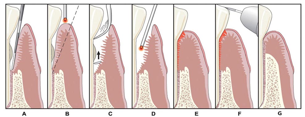
a) Measurement of pockets Your doctor uses a probe to measure the depth of the periodontal pockets. This measurement provides key information about how much the gum has detached from the root.
b) Laser removal of bacteria The PerioLase® MVP-7™ uses pulsed light to dissolve diseased tissue within the pocket and kills the living bacteria that cause gum disease. The laser’s light is not absorbed by water so the surrounding tissue, root, and bone remain unaffected by the laser. Surgery is far less invasive and more comfortable than other procedures.
c) Removal of calculus Calculus is hardened plaque that has attached to your teeth and bone. Ultrasonic scalers scrape away the calculus that has formed in pockets beneath the gum line. Although the laser cannot completely remove the calculus in the earlier steps, it assists by denaturing the calculus, breaking down its chemical make-up, and making it easier to remove.
d) Clot Formation The laser is used a second time at the bottom of the pockets to stimulate the soft tissue, root, and bone to incite regeneration and attachment. Simultaneously the tissue becomes sticky and begins forming a sealed clot around the tooth root. This clot creates a clean, stable environment to foster fast and complete healing.
e) Tissue compression Gum tissue is compressed against the tooth’s root surface to promote reattachment and eliminate the need for painful sutures.
f) Bite adjustment Your doctor adjusts your bite to minimize trauma on the teeth, particularly in areas where bone loss has occurred.
Download the LANAP Laser Pack
Next LANAP vs. Traditional Gum Surgery >>